Home

ricordi Hubert Hudson Mamma caudal vena cava dog Giocoso facile da gestire Di

CT variants of the caudal vena cava in 121 small breed dogs - Ryu - 2019 -  Veterinary Radiology & Ultrasound - Wiley Online Library
CT variants of the caudal vena cava in 121 small breed dogs - Ryu - 2019 - Veterinary Radiology & Ultrasound - Wiley Online Library
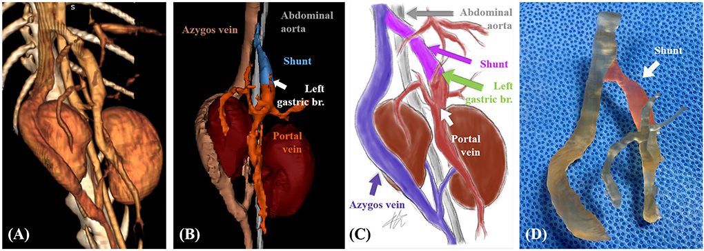
Frontiers | Case report: Application of three-dimensional technologies for  surgical treatment of portosystemic shunt with segmental caudal vena cava  aplasia in two dogs
Frontiers | Case report: Application of three-dimensional technologies for surgical treatment of portosystemic shunt with segmental caudal vena cava aplasia in two dogs
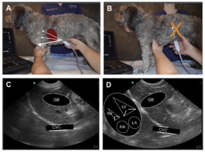
ECC and IM Blog, March 2022 - The Characterization of the Caudal Vena Cava  and Hepatic Veins - FASTVet
ECC and IM Blog, March 2022 - The Characterization of the Caudal Vena Cava and Hepatic Veins - FASTVet

FIGURE 1. Anatomy of the canine liver (caudal or visceral aspect), with the  lobes, porta hepatis, caudal vena cava… | Anatomy organs, Ultrasound,  Diagnostic imaging
FIGURE 1. Anatomy of the canine liver (caudal or visceral aspect), with the lobes, porta hepatis, caudal vena cava… | Anatomy organs, Ultrasound, Diagnostic imaging

Vet Emergency Ultrasound: Interpretation of the Caudal Vena Cava - YouTube

Measurement of the CVC amin-HV-B , Ao min-HV-B (HV B-Mode). CVC, caudal...  | Download Scientific Diagram
Measurement of the CVC amin-HV-B , Ao min-HV-B (HV B-Mode). CVC, caudal... | Download Scientific Diagram
Dog - Abdomen - Pelvis (CT): normal anatomy | vet-Anatomy

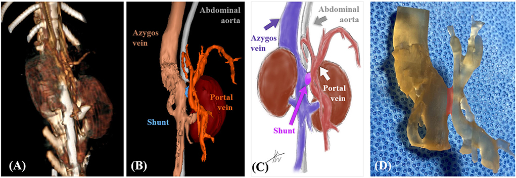
Frontiers | Case report: Application of three-dimensional technologies for  surgical treatment of portosystemic shunt with segmental caudal vena cava  aplasia in two dogs
Frontiers | Case report: Application of three-dimensional technologies for surgical treatment of portosystemic shunt with segmental caudal vena cava aplasia in two dogs

Caudal vena cava to aorta ratio of dogs with spontaneous circulatory shock  | Veterinary 33
Caudal vena cava to aorta ratio of dogs with spontaneous circulatory shock | Veterinary 33

Dog 1 of PH group; APSS from portal vein to cranial vena cava via left... |  Download Scientific Diagram
Dog 1 of PH group; APSS from portal vein to cranial vena cava via left... | Download Scientific Diagram

Post-contrast CT images of severe caudal vena cava obstruction in dog... |  Download Scientific Diagram
Post-contrast CT images of severe caudal vena cava obstruction in dog... | Download Scientific Diagram

CT variants of the caudal vena cava in 121 small breed dogs - Ryu - 2019 -  Veterinary Radiology & Ultrasound - Wiley Online Library
CT variants of the caudal vena cava in 121 small breed dogs - Ryu - 2019 - Veterinary Radiology & Ultrasound - Wiley Online Library

MULTIDETECTOR ROW COMPUTED TOMOGRAPHY AND ULTRASOUND CHARACTERISTICS OF CAUDAL  VENA CAVA DUPLICATION IN DOGS - Bertolini - 2014 - Veterinary Radiology &  Ultrasound - Wiley Online Library
MULTIDETECTOR ROW COMPUTED TOMOGRAPHY AND ULTRASOUND CHARACTERISTICS OF CAUDAL VENA CAVA DUPLICATION IN DOGS - Bertolini - 2014 - Veterinary Radiology & Ultrasound - Wiley Online Library

Figure 1 from Computed tomographic characteristics of collateral venous  pathways in dogs with caudal vena cava obstruction. | Semantic Scholar
Figure 1 from Computed tomographic characteristics of collateral venous pathways in dogs with caudal vena cava obstruction. | Semantic Scholar

Radiographic evaluation of caudal vena cava and descending aorta in  indigenous dog breeds of Tamil Nadu
Radiographic evaluation of caudal vena cava and descending aorta in indigenous dog breeds of Tamil Nadu

Cardiovascular System – Canine Anatomy for Beginners

Ultrasonography of the Abdominal Vasculature | Today's Veterinary Practice

Caudal vena cava thrombosis in two dogs - Small Animal Ultrasonography

Canine Venous Return Caudal Vena Cava Diagram | Quizlet

Caudal vena cava - vet-Anatomy - IMAIOS

Caudal vena cava point-of-care ultrasound in dogs with degenerative mitral  valve disease without clinically important right heart disease -  ScienceDirect
Caudal vena cava point-of-care ultrasound in dogs with degenerative mitral valve disease without clinically important right heart disease - ScienceDirect

Segmental aplasia of the caudal vena cava in a dog. | Semantic Scholar

Figure 5 from Computed tomographic characteristics of collateral venous  pathways in dogs with caudal vena cava obstruction. | Semantic Scholar
Figure 5 from Computed tomographic characteristics of collateral venous pathways in dogs with caudal vena cava obstruction. | Semantic Scholar